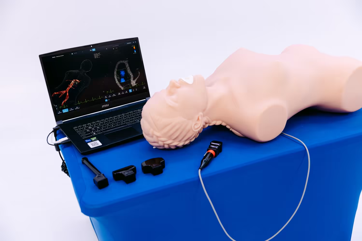
The rapid integration of Point-of-Care Ultrasound (POCUS) across modern healthcare systems has permanently transformed diagnostic medicine. Once confined strictly to the radiology department, bedside sonography is now an essential clinical tool in emergency medicine, intensive care, obstetrics, and cardiology. However, mastering the unique psychomotor skills and cognitive spatial awareness required to interpret dynamic, artifacts-laden acoustic images presents an exceptionally steep learning curve. To address this educational challenge, medical universities and teaching hospitals globally rely heavily on simulation-based education.
As the market for educational medical devices expands, conducting a thorough ultrasound simulator comparison becomes an absolute necessity for institutional procurement committees. Selecting the right ultrasound simulator requires a deep understanding of how different technological architectures translate to actual clinical proficiency. A comprehensive review of available systems reveals that an optimal ultrasound simulation platform must balance physical fidelity, pathological diversity, and objective metrics tracking to effectively elevate student practice.
Technological Architectures in Sonography Simulation
To establish a baseline for an effective ultrasound simulator comparison, it is essential to first analyze the underlying technologies that drive modern sonographic simulation. Manufacturers generally develop an ultrasound simulator using one of two primary architectural frameworks: physical tissue-equivalent phantoms or fully synthetic virtual reality (VR) environments.

Physical Tissue-Equivalent Phantoms
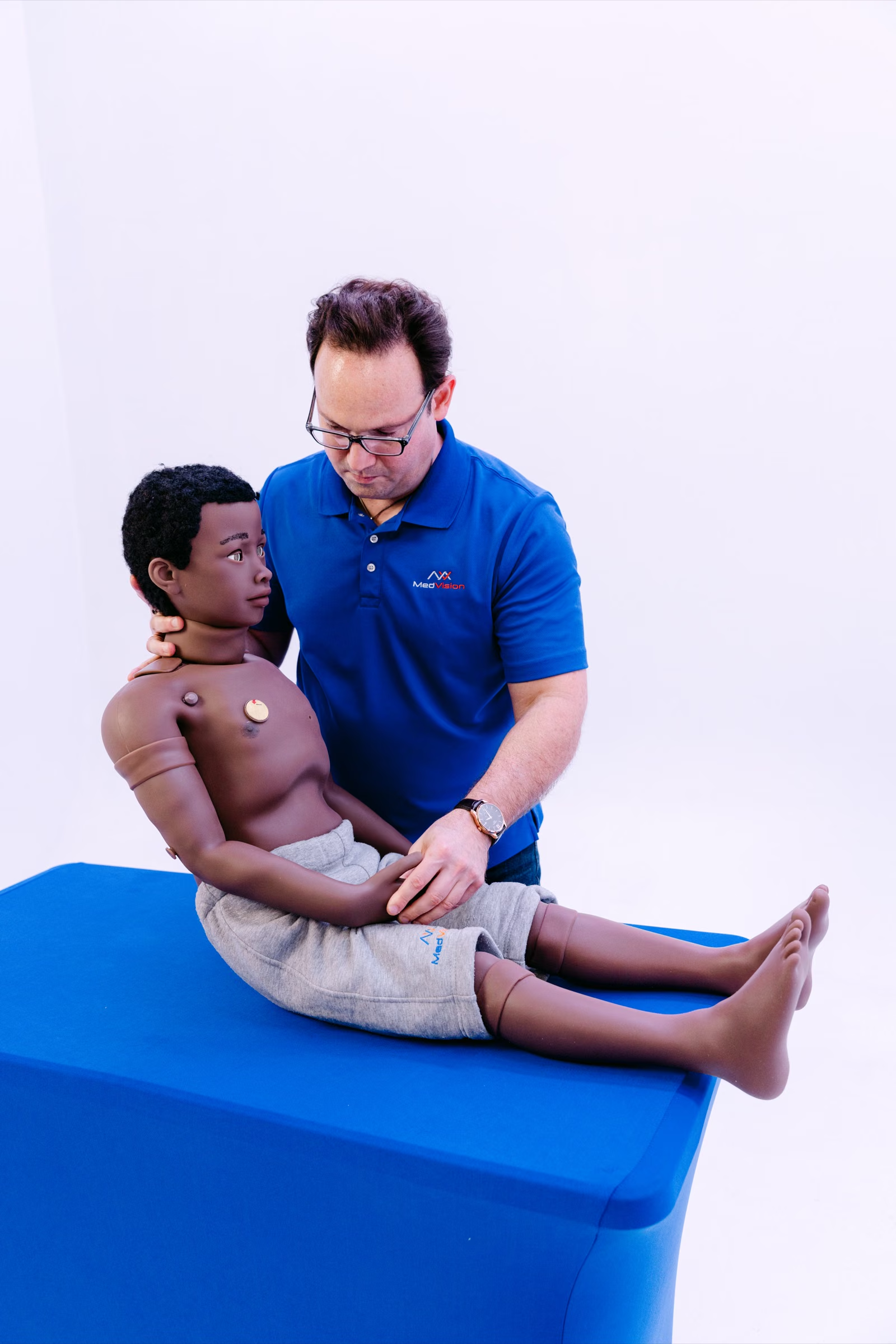
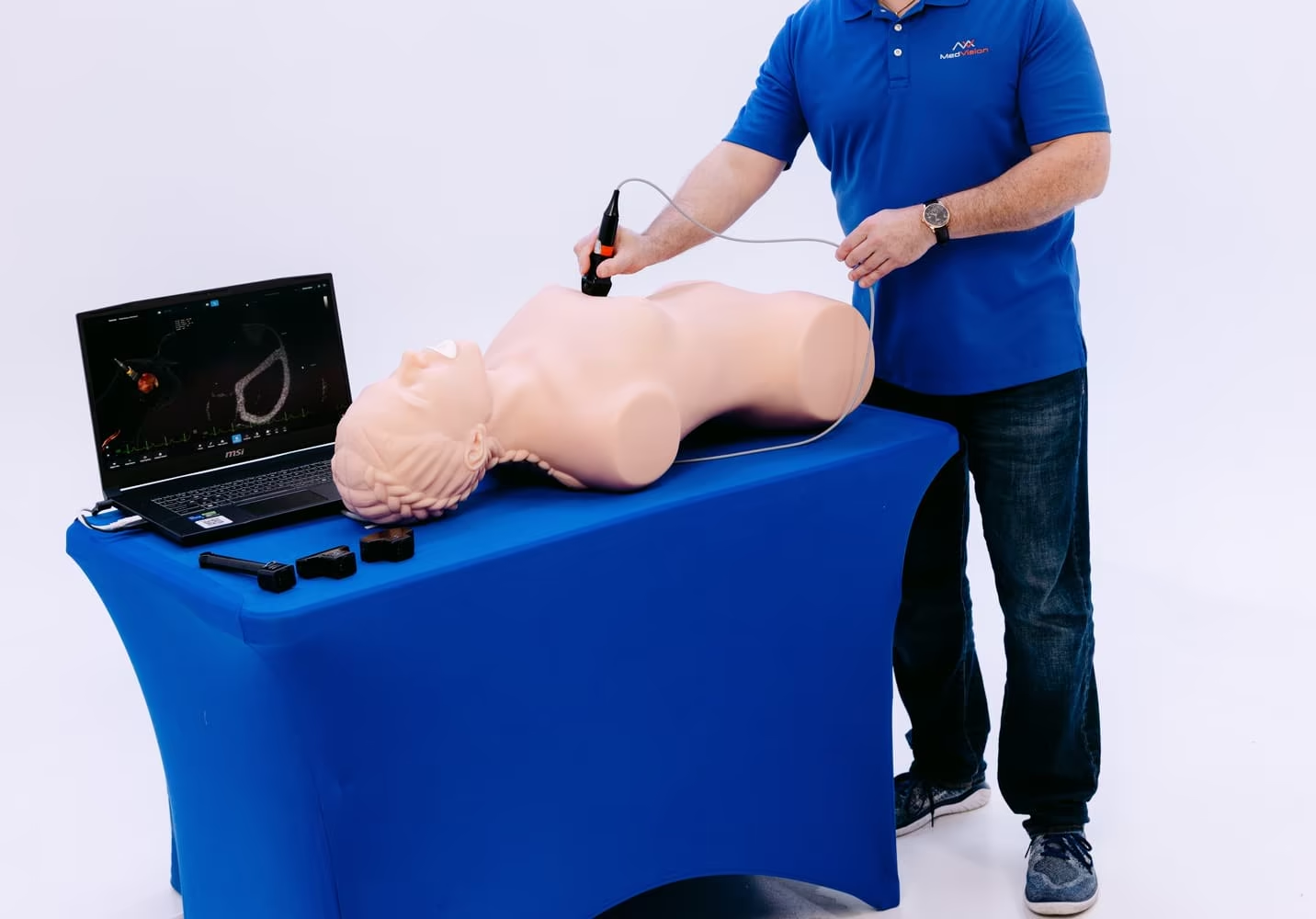
Physical phantoms are engineered from specialized, proprietary polymers that perfectly mimic the acoustic impedance, attenuation, and scattering properties of human flesh, fat, fluid, and bone. When medical students utilize this type of ultrasound model, they connect a real, clinical-grade ultrasound machine to the phantom. As they apply actual acoustic gel and manipulate the live transducer, the machine processes the sound waves to generate a genuine, real-time ultrasound image on the monitor.
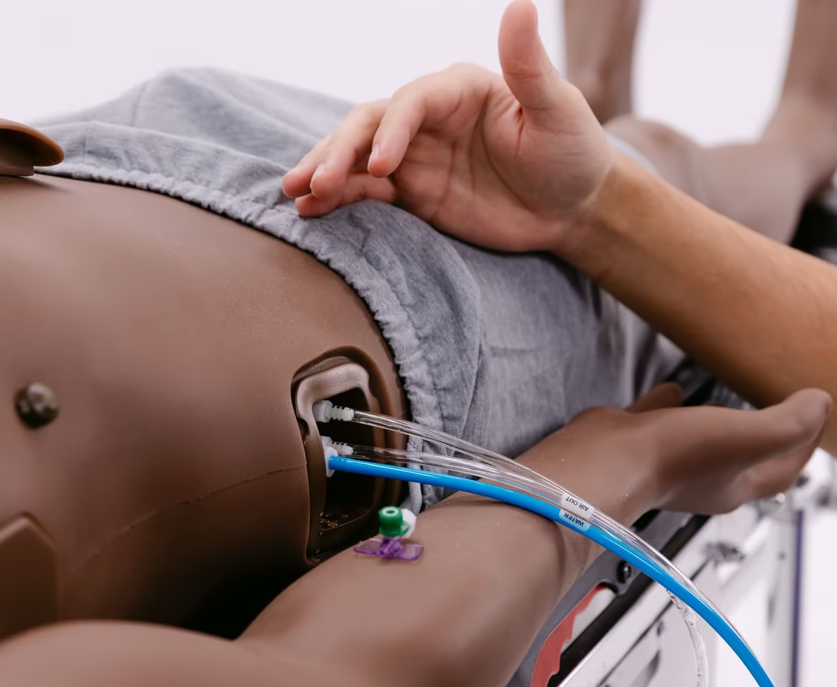
The primary advantage of this physical approach is its absolute tactile and mechanical authenticity. The learner experiences the exact physical resistance of scanning across human ribs or compressing a deep vein. This makes physical phantoms an excellent choice for mastering basic psychomotor skills, needle guidance, and interventional procedures like ultrasound-guided central line placement. However, physical phantoms are structurally static; a single phantom cannot dynamically transition from representing a healthy liver to displaying a massive hepatic abscess, significantly limiting case diversity.
Virtual Reality and Computer-Based Simulation
Conversely, a virtual reality ultrasound simulator replaces the real imaging machine with an entirely digital, algorithmic computing system. The trainee uses a mock, sensor-tracked transducer probe over a generalized, non-acoustic mannequin or a tracking pad. The internal simulation software holds an extensive cloud-based repository of authentic, pre-recorded patient datasets. As the user moves the virtual probe, the computer instantly calculates the 3D spatial coordinates and projects the corresponding cross-sectional ultrasound slice onto the screen.
The defining benefit of a digital ultrasound simulator is its near-infinite pathological variability. With a few clicks on the user interface, an instructor can alter the virtual patient's anatomy from a normal physiological state to a rare, acute life-threatening crisis. This software-based versatility forms the core of modern diagnostic learning. Furthermore, digital systems automatically track performance metrics - such as probe angulation accuracy, organ visualization time, and diagnostic precision - providing completely objective data to evaluate student practice. The limitation, however, is that virtual reality systems rely on simulated haptics, which can occasionally lack the raw, organic mechanical feedback of an actual physical phantom.
Critical Evaluation Metrics for Institutional Selection
When performing an institutional ultrasound simulator comparison, academic directors must look beyond marketing descriptions and rigorously evaluate specific educational metrics. An optimized ultrasound simulator must satisfy three critical pillars: haptic fidelity, pathological case breadth, and pedagogical autonomy.
Haptic Fidelity and Spatial Tracking
The core of sonographic technique relies on fine-motor manipulation: sliding, rocking, rotating, and fanning the probe. Therefore, any premium ultrasound simulator must possess ultra-high-resolution spatial tracking technology. If the digital simulation software exhibits any mechanical lag or micro-latency between the physical movement of the mock probe and the rendering of the ultrasound image on the monitor, the training becomes actively detrimental. Inaccurate tracking can inadvertently teach students incorrect muscle memory, which can negatively impact their real-world clinical performance. High-fidelity magnetic or optical tracking systems ensure that the digital rendering perfectly replicates real-world physics.

Pathological Database Breadth
A truly effective diagnostic ultrasound simulator must expose learners to a vast spectrum of clinical conditions that they might not encounter for years during standard clinical rotations. When conducting an ultrasound simulator comparison, institutions should carefully evaluate the depth of the software's clinical library. A premium platform should include extensive, validated datasets covering congenital heart defects, various stages of ectopic pregnancies, advanced abdominal trauma, and complex vascular pathologies. The ability to seamlessly layer multiple pathologies onto a single virtual patient model dramatically enhances cognitive flexibility and diagnostic competence.
Comprehensive Comparative Table: Physical vs. Virtual Simulation
To effectively summarize this comprehensive ultrasound simulator comparison, the following table delineates the core operational differences, pedagogical advantages, and inherent limitations of the two primary modalities utilized in modern sonographic education.
Emergency Medicine and Critical Care Simulation
In the high-stakes, chaotic environment of the emergency department and intensive care unit, rapid diagnostic imaging is a matter of life and death. Consequently, evaluating the trauma capabilities of a system is a mandatory component of any thorough ultrasound simulator comparison. Emergency physicians must routinely perform the FAST (Focused Assessment with Sonography for Trauma) examination to rapidly detect internal hemorrhage.
When a residency program invests in an advanced ultrasound simulator, trainees can rigorously practice this exact protocol in a perfectly controlled, risk-free environment. During an acute trauma simulation, the instructor can program the ultrasound software to instantly display massive free fluid in Morison’s pouch. If the resident fails to angle the mock probe correctly on the simulator, the internal bleeding goes entirely undetected. This intense, scenario-based practice ensures that when the learner eventually treats a live trauma patient, their spatial awareness and sonographic reflexes are flawless. Furthermore, critical care simulation heavily utilizes the ultrasound simulator to teach the RUSH (Rapid Ultrasound in Shock) protocol, allowing students to instantly evaluate a virtual patient's cardiac output and intravascular volume status using simulated acoustic windows.
Obstetrics, Gynecology, and Transvaginal Simulation
The fields of obstetrics and gynecology present incredibly unique challenges that demand specialized ultrasound simulation technology. Performing a transvaginal ultrasound is an intimate, highly invasive procedure that requires immense physical sensitivity and profound anatomical knowledge. Relying on live human patients for the initial stages of transvaginal learning is ethically problematic and highly uncomfortable. Therefore, deploying a dedicated pelvic ultrasound simulator is absolutely essential for safe, compassionate medical training.
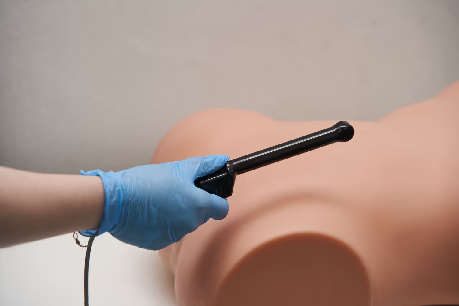
A premium transvaginal ultrasound simulator provides an anatomically accurate pelvic mannequin combined with high-fidelity digital rendering. During this highly specialized simulation, the trainee navigates the virtual probe to assess ovarian follicles, measure nuchal translucency, and detect severe congenital fetal abnormalities. An exhaustive ultrasound simulator comparison will reveal that the best platforms offer dynamic fetal simulation, where the virtual fetus physically moves, forcing the student to continuously adjust their sonographic plane. This rigorous practice builds the necessary muscle memory and cognitive resilience required for high-level maternal-fetal healthcare.
Artificial Intelligence Integration in Ultrasound Simulation
As we look toward the immediate future of medical education, the integration of Artificial Intelligence (AI) is completely redefining the capabilities of the modern ultrasound simulator. Advanced simulation platforms now incorporate highly sophisticated machine learning algorithms that function as autonomous digital instructors.
During a practice session, the AI-enhanced ultrasound system continuously analyzes the student's physical probe angulation and digital image acquisition in real-time. If a learner is struggling to locate the gallbladder, the ultrasound simulator proactively overlays visual directional arrows onto the screen, guiding the trainee’s hand toward the correct acoustic window. This predictive, algorithmic guidance maximizes the efficiency of independent learning. A detailed ultrasound simulator comparison confirms that platforms utilizing AI-driven simulation dramatically reduce the logistical burden on human faculty, allowing students to log countless hours of objective, metric-driven practice outside of standard laboratory hours.
The Economic ROI of an Ultrasound Simulator
Acquiring state-of-the-art laboratory equipment for immersive ultrasound simulation requires a formidable institutional capital expenditure. High-fidelity VR computing systems and highly durable physical phantom models are undeniably expensive. However, a comprehensive financial review definitively proves that the Return on Investment (ROI) for a modern ultrasound simulator is exceptionally robust.
First, utilizing an ultrasound simulator for foundational skills training drastically reduces the daily mechanical wear and tear on an expensive, clinical-grade hospital ultrasound machine. High-frequency transducers are fragile and easily broken by inexperienced novices; keeping them safely in the clinical ward while students use the simulator saves thousands of dollars in replacement costs. More importantly, rigorous simulation training massively mitigates severe institutional liability. A missed sonographic diagnosis in the emergency department can result in catastrophic patient outcomes and multi-million dollar malpractice litigation. By mandating that all residents achieve a mathematically verified baseline of diagnostic competency on an ultrasound simulator before treating live humans, the hospital legally and ethically safeguards its patients. The immense financial resources saved by preventing just one major clinical error effortlessly justify the entire operational cost of the simulation laboratory.
Conclusion
In final analysis, the monumental shift toward competency-based medical education requires the widespread, global integration of advanced diagnostic technologies. Conducting a rigorous, evidence-based ultrasound simulator comparison empowers curriculum directors to select the optimal educational tools for their unique clinical cohorts. Whether an institution prioritizes the absolute tactile authenticity of physical tissue phantoms for interventional skills, or the immense pathological diversity of a virtual reality ultrasound simulator for diagnostic mastery, the ultimate goal remains universally identical: supreme patient safety.
By strategically implementing comprehensive ultrasound simulation programs, medical universities establish an incredibly powerful, objective, and psychologically safe learning environment. This technology-enhanced practice ensures that every single graduating physician possesses the deeply refined spatial awareness, flawless probe mechanics, and impeccable diagnostic accuracy necessary to succeed. Ultimately, heavy institutional investment in the modern ultrasound simulator guarantees that when these highly trained professionals finally face critical medical emergencies in the real world, their sonographic response will be flawlessly swift, fiercely accurate, and profoundly life-saving.
References
- Ahmad, R., et al. (2020). "The role of high-fidelity ultrasound simulation in medical education: A systematic review." Journal of Ultrasound in Medicine, 39(12), 2285-2296.
- Gottlieb, M., & Bailitz, J. (2018). "Point-of-care ultrasound training in emergency medicine: The impact of simulation models." Annals of Emergency Medicine, 72(4), 450-457.
- Kaufman, J. (2019). "Cognitive load and simulation in healthcare: Optimizing the learning environment for medical students." Medical Education Online, 24(1), 162-171.
- Neff, L., et al. (2021). "Virtual reality and physical phantom models in obstetrics ultrasound training." Ultrasound in Obstetrics & Gynecology, 58(2), 211-219.
- Ziv, A., Rootenberg, L., & Macrae, H. (2016). "Simulation-based training in healthcare: A critical evaluation of the evidence base." Quality and Safety in Health Care, 15(suppl 1), i34-i43.
FAQ
The primary purpose of an ultrasound simulator comparison is to meticulously evaluate the technological differences between physical tissue phantoms and virtual reality systems. This review allows healthcare institutions to select the exact ultrasound simulator that perfectly aligns with their specific educational curriculum, ensuring students acquire both mechanical skills and diagnostic cognitive abilities.
An ultrasound simulator dramatically improves emergency practice by providing a completely safe environment to rehearse life-saving protocols like the FAST and RUSH exams. Through rigorous ultrasound simulation, trainees build profound muscle memory, allowing them to instantly identify lethal conditions - such as internal bleeding or cardiac tamponade - without jeopardizing live patient safety.
While an advanced ultrasound simulator cannot entirely replace the organic variability of scanning a live human on a real ultrasound machine, it perfectly bridges the critical gap between textbook theory and clinical reality. The simulation phase safely eliminates the steepest, most dangerous part of the learning curve, ensuring students possess a solid baseline of sonographic skills before touching a real patient.
Transvaginal ultrasound is an intimate, invasive procedure. Utilizing a dedicated pelvic ultrasound simulator allows residents to repeatedly practice their probe mechanics and spatial orientation on a highly accurate physical mannequin. This simulation ensures they can compassionately and accurately diagnose fetal anomalies without causing physical or emotional discomfort to live women during early educational practice.
Virtual demo
See it on a real simulator
A MedVision specialist shows the simulator live and answers your questions. Remote setup included · Online Training Included.
Request a virtual demo